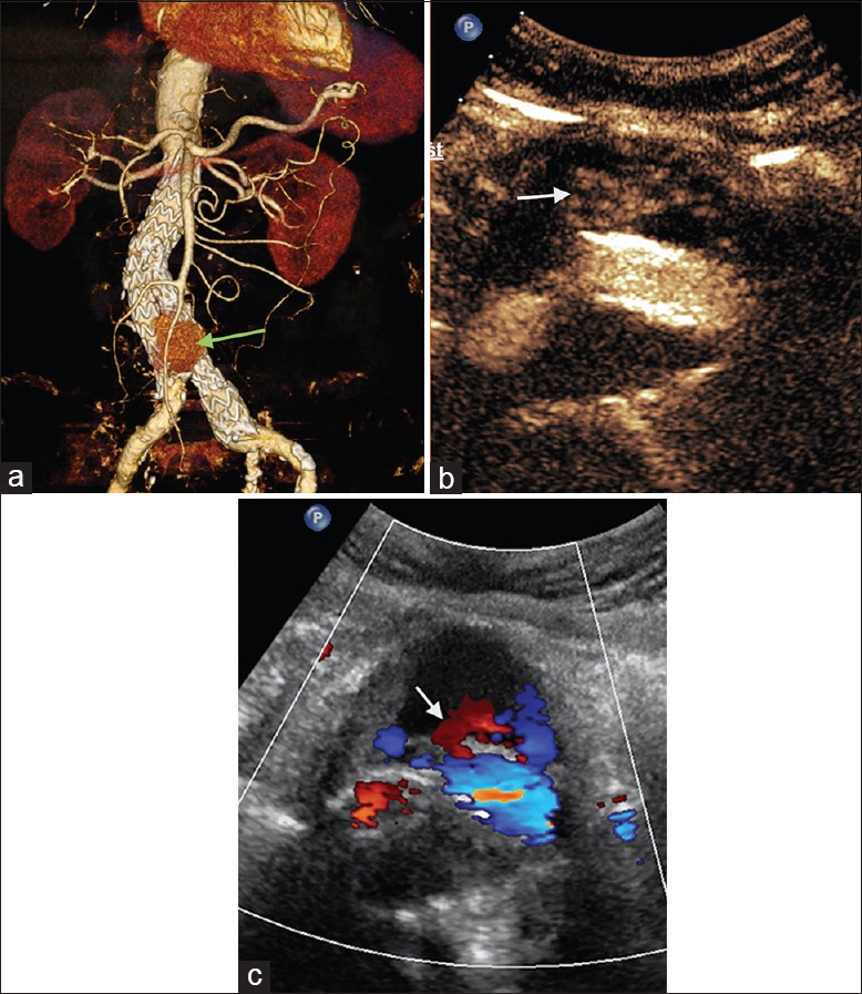
Figure 2.

A 83-year-old male patient, postendovascular aneurysm repair. (a) Computed tomography angiography shows contrast agent around the stent with obscure leak site (green arrow). (b) Contrast-enhanced ultrasound shows leak at the lower part of the stent's right leg (type Ib) (white arrow). (c) Color Doppler flow imaging shows outflow from the stent (white arrow).
