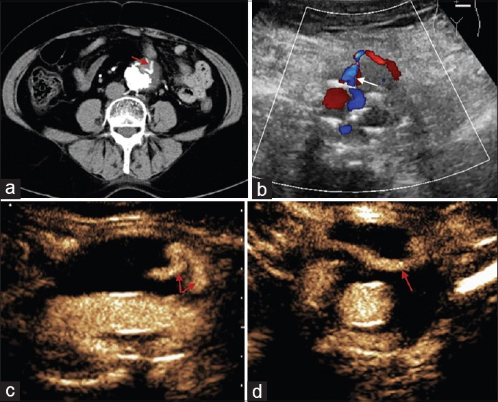
Figure 3.

A 71-year-old female patient, postendovascular aneurysm repair. (a) Computed tomography angiography shows an endoleak in an aneurysm (red arrow). (b) Color Doppler flow imaging shows the leak's origin from an inferior mesenteric artery (white arrow). (c) Contrast-enhanced ultrasound demonstrates two reflux flow (red arrows), and (d) the flow unseen by color Doppler flow imaging and computed tomography angiography travels horizontally (red arrow).
