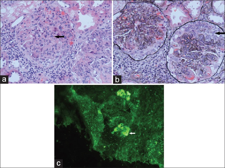
Figure 1.

Renal pathology of the patient. (a) H and E staining (original magnification, ×400) of renal biopsy demonstrating proliferation of mesangial cells and matrix (arrow). (b) Periodic acid-silver metheramine staining (original magnification, ×400) of renal biopsy showed cellular crescents (arrow). (c) Immunofluorescence of renal biopsy showed immunoglobulin A deposits in the mesangium (arrow).
