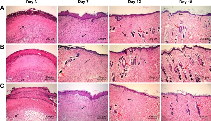
Figure 2.
Wound healing histology for each group at days 3, 7, 12, and 18 after surgery.
Notes: HE staining shows collagen fibers stained pale pink, cytoplasm stained purple, nuclei stained blue, and red blood cells stained cherry red. A, B, and C show gauze group, PVA/COS-AgNPs nanofiber group, PVA/COS-AgNPs nanofiber plus SB431542 group, respectively. The black and red arrows indicate blood vessels and hair follicles, respectively.
Abbreviation: HE, hematoxylin–eosin.

