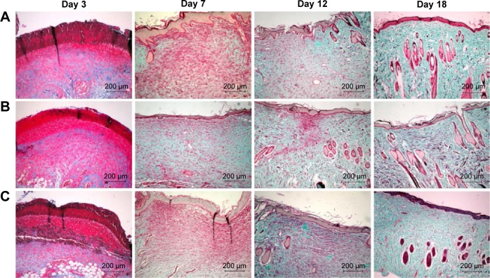
Figure 3.
Masson’s trichome staining of biopsies of rat skin tissue.
Notes: Collagen is stained blue-green, while cytoplasm, red blood cells, and nuclei are stained red. Masson-trichrome staining of collagen at day 3, 7, 12, 18 post-wounding: (A) gauze, (B) PVA/COS-AgNPs nanofiber, (C) PVA/COS-AgNPs nanofiber plus SB431542.

