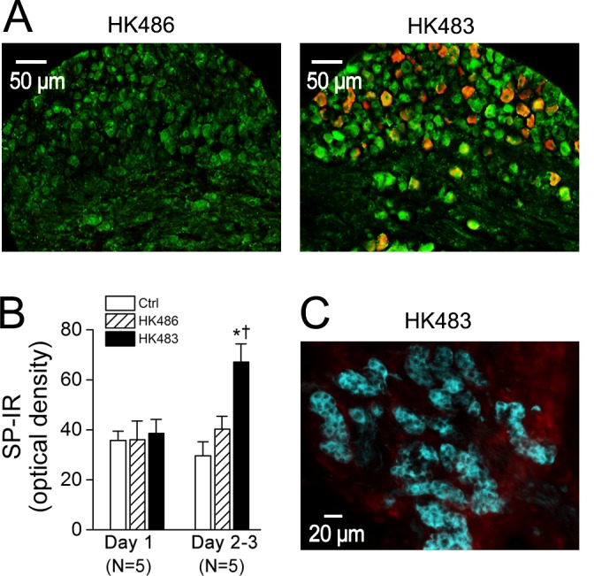
Fig 8. Expression of nucleoprotein-immunoreactivity (NP-IR, orange) in substance P immunoreactivity (SP-IR) neurons (green) in the nodose ganglion and tyrosine hydroxylase immunoreactivity (TH-IR) neurons (cyan) in the carotid body.

A: At 2 dpi, nodose ganglion NP-IR appears in a HK483 (right) but not HK486 mice (left) and HK483 virus increases the density of SP-IR in the nodose ganglionic neurons. In contrast, NP-IR is undetectable in the nodose ganglion of infected HK483 and HK486 mice at 1 dpi (data not shown). B: The density of SP-IR in the nodose ganglion is not significantly changed by HK483 virus until 2 dpi. Because of the similarity of the SP-IR data at 2 and 3 dpi (N = 3 and 2, respectively) in either HK483 (64 ± 11 vs. 69 ± 15) and HK486 mice (40 ± 9 vs. 41 ± 11), they were grouped. C: No viral replication in the carotid body of a HK483 mouse in which glomus cells are marked by TH-IR (cyan) at 3 dpi in a HK483 mice. N = 5 for each group; * and † P < 0.05 compared HK483 data to Ctrl and HK486 data respectively.
