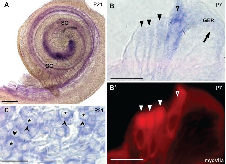
Fig 4. In situ hybridization probes against caldendrin, CaBP1-L, and CaBP1-S label hair cells and the spiral ganglion.
The pan-CaBP1 probe was designed to recognize the 3’ end of the Cabp1 gene such that it should recognize all three variants of CaBP1. (A) In situ hybridization of a P21 mouse cochlea, showing pan-CaBP1 labeling in the spiral ganglion (SG) and the organ of Corti (OC). (B-B’) 10 μm-thick plastic section showing in situ hybridization signal in the organ of Corti (P7) double labeled with antibodies against myosin VIIa. Open triangles indicate inner hair cells; filled triangles indicate outer hair cells; arrow indicates the inner supporting cells in greater epithelial ridge (GER). (C) 10 μm-thick plastic section showing expression of CaBP1/caldendrin in situ hybridization in the spiral ganglion. Asterisks indicate spiral ganglion neuron cell bodies; arrowheads indicate pan-CaBP1 signal in cells surrounding neuron cell bodies. Scale bars: 200 μm (A); 20 μm (B, C).

