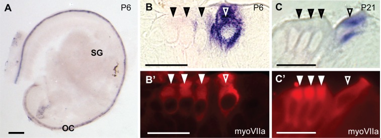
Fig 5. In situ hybridization probes against CaBP2-alt label cochlear hair cells.
The RNA probe was designed to recognize exon 1a unique to the CaBP2-alt splice variant. (A) CaBP2-alt in situ hybridization signal in a mouse cochlea at P6. (B-C’) 10 μm-thick plastic sections of mouse cochlea at P6 (B) or P21 (C) were hybridized with CaBP2-alt in situ probe and labeled with antibodies against myosin VIIa (myoVIIa) for immunofluorescence. CaBP2-alt in situ hybridization signal (B,C) is localized to myoVIIa-labeled inner hair cells (B’, C’). Filled triangles indicate outer hair cells; open triangles indicate inner hair cells. Scale bars: 200 μm (A); 20 μm (B-C’). OC, organ of Corti; SG, spiral ganglion.

