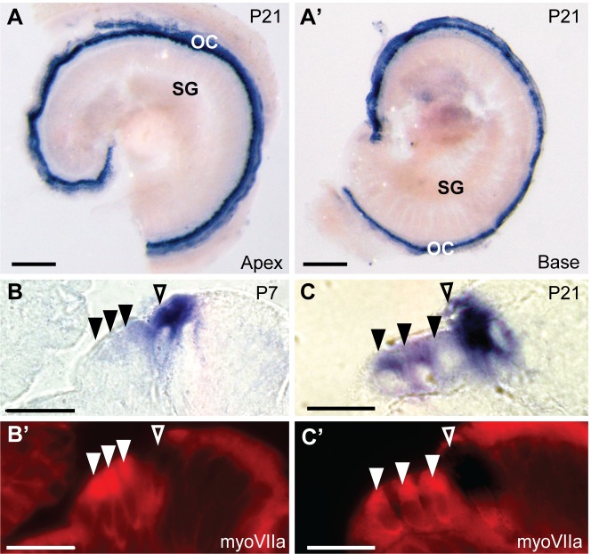
Fig 6. In situ hybridization probes against CaBP1-alt, CaBP2-L and CaBP2-S label inner and outer hair cells.
The RNA probe was designed to recognize all splice variants of CaBP2. (A-A’) In situ hybridization signal in the apical (A) and basal (A’) turn of a mouse cochlea at P21. (B-C’) 10 μm-thick plastic sections of cochlea hybridized with pan-CaBP2 in situ probe and immunofluorescently labeled with antibodies against myosin VIIa (myoVIIa) immunofluorescence at P7 (B) or P21 (C). Images represent in situ hybridization signal (A,A’, B,B’) or myoVIIa immunofluorescence (B’,C’). Filled triangles indicate outer hair cells; open triangles indicate inner hair cells. Scale bars: 200 μm (A-A’), 20 μm (B-C’). OC, organ of Corti; SG, spiral ganglion.

