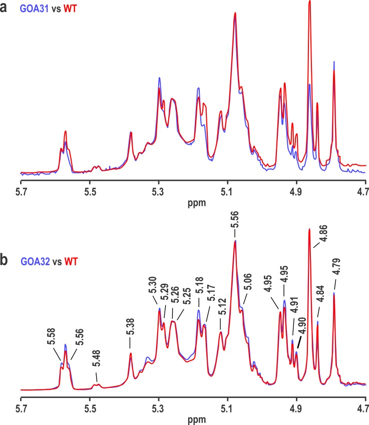
Fig 5. NMR reveals a reduction in β-1,2 linked mannan (peak 4.86) of GOA31.
The 600 MHz proton NMR spectra of mutants (in blue) GOA31 (top) and GOA32 (bottom) are compared to the NMR spectrum of WT (in red) by overlaying the spectra and adjusting the resonance at 5.08 ppm to the same height in each pair of spectra. A prominent loss of the 4.86 ppm resonance (β-1,2 mannan) occurred in GOA32.

