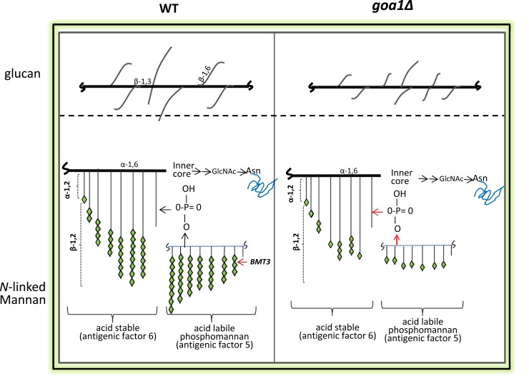
Fig 6. Models of the cell wall polysaccharides and linkages are shown for WT (left) and goa1∆ (GOA31, right).
Our data predict that GOA31 β-1,6 glucan is more highly branched but with shorter side chains compared to WT cells (upper). For N-linked mannan, β-linked mannans, particularly of acid-labile phosphomannan, are truncated in GOA31 compared to WT cells due to defects in β-1,2 mannan elongation.

