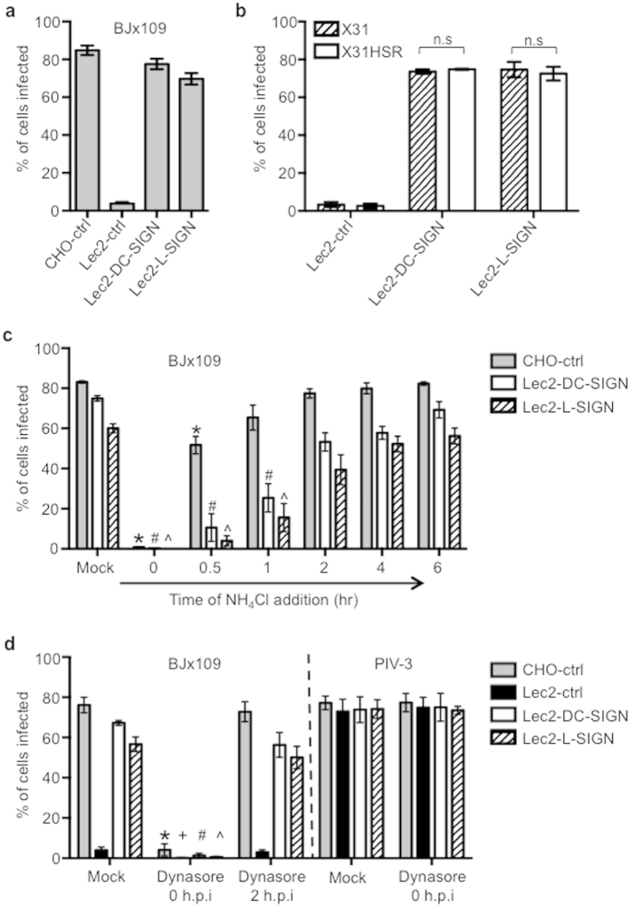
Figure 1. Infectious entry of IAV into SIA-deficient Lec2 cells expressing DC-SIGN/L-SIGN.
(a,b) CHO-ctrl, Lec2-ctrl, Lec2-DC-SIGN or Lec2-L-SIGN cells were infected with (a) BJx109 or (b) HKx31 and HKx31 HSR (MOI 50 PFU/cell) for 8 hr at 37 °C. Data represent the mean percent infected cells (±1 SEM) from at least 4 independent fields and are representative of multiple independent experiments. (n.s = no significant difference for HKx31 compared to HKx31 HSR (Lec2-DC-SIGN; P = 0.37, Lec2-L-SIGN; P = 0.71) (Student’s t-test; two-tailed)). (c) CHO-ctrl, Lec2-DC-SIGN and Lec2-L-SIGN cells were incubated with BJx109 (MOI 50 PFU/cell) for 60 min at 37 °C and then washed and cultured 8 hr (mock). In some wells, 10 mM NH4Cl was added to the cells at the time of virus addition (t = 0 hr) or at various times thereafter to prevent further infection. All cells were incubated for 8 hr. Data represent the mean percent infected cells (±1 SEM). *#^significantly different compared to mock for CHO-ctrl, Lec2-DC-SIGN and Lec2-L-SIGN cells, respectively. p < 0.05, one-way ANOVA followed by Tukey’s post-hoc test. (d) CHO-ctrl, Lec2-ctrl, Lec2-DC-SIGN and Lec2-L-SIGN cells were incubated with 107 PFU of BJx109 (MOI 50 PFU/cell) for 60 min at 37 °C and then washed (mock). In some wells, 50 μM of dynasore was added to the cells at the time of infection (t = 0 hr) and replaced after washing, or dynasore was added two hours after incubation with virus (t = 2 hr). Cells were incubated for a total of 8 hr. Additional wells incubated with 106 FFU of PIV-3 (MOI 5 FFU/cell) in the presence or absence of 50 μM dynasore were fixed and stained for expression of PIV-3 NP at 16 hr post-infection. Data represent the mean percent infected cells (±1 SEM). *+#^significantly different compared to mock for CHO-ctrl, Lec2-ctrl, Lec2-DC-SIGN and Lec2-L-SIGN cells, respectively. p < 0.05, one-way ANOVA followed by Tukey’s post-hoc test. Data in (c,d) are pooled from 3 independent experiments.

