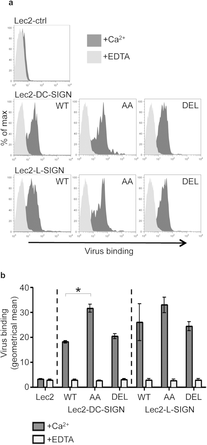
Figure 3. IAV binding to cells expressing WT, AA or DEL mutants of DC-SIGN/L-SIGN.

(a) IAV binding to Lec2 cells expressing -WT, -AA or -DEL mutants of DC-SIGN/L-SIGN is Ca2+-dependent. Representative histograms show cell-surface binding of biotinylated IAV (strain BJx109) to Lec2 cells expressing -WT, -AA or -DEL forms of DC-SIGN/L-SIGN in the presence of 10 mM CaCl2 or 5 mM EDTA. No binding to Lec2-ctrl cells was observed. (b) IAV binding is presented as the geometrical mean of the fluorescence intensity (±1 SEM) from FACS histograms of triplicate samples. (*virus binding to Lec2-DC-SIGN-WT cells was significantly different compared with Lec2-DC-SIGN-AA cells. p < 0.05, One way ANOVA, followed by Tukey’s post-hoc test.)
