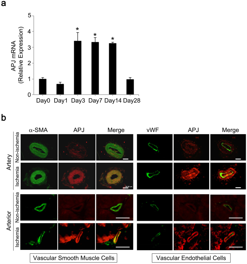
Figure 5. APJ expression in vascular smooth muscle cells of ischemic arteries.
(a) Quantitative RT-PCR for APJ gene expression was performed in ischemic arteries from mouse hindlimbs at day 0 (pre-ligation), 1, 3, 7, 14, and 28 post-ligation. APJ mRNA level in ligated arteries was significantly increased between days 3 and 14 post-ligation and returned to baseline level at day 28 post-ligation. *p < 0.01 vs. baseline (day 0). (b) Vascular smooth muscle cells, but not endothelial cells, expressed APJ in ischemic arteries. At day 14, ligated arteries were collected and immunohistochemistry for APJ (red), vWF (green), and α-SMA (green) was performed to identify cells expressing APJ in ischemic arteries. Scale bar = 50 μm.

