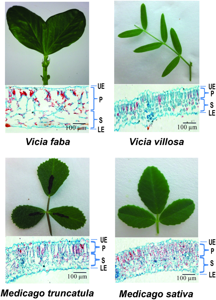
Figure 4. The leaf microstructure of Vicia faba, Vicia villosa, Medicago truncatula, and Medicago sativa.
Cross sections of leaves were stained in 0.05% aqueous safranin and counterstained with an alcoholic solution of fast green FCF. P, palisade tissue. S, spongy tissue. UE, upper epidermis. LE, lower epidermis.

