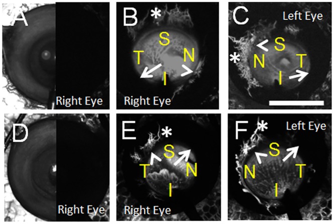
Fig 7. Aqueous Angiography in Enucleated Human Eyes.

Aqueous angiography was performed on enucleated eyes from two female subjects not known to have glaucoma at 10 mm Hg (subject 1 = (A-C) and subject 2 = (D-F)). Both right and left eyes from each subject were investigated and shown at 10–25 seconds. (A/D) Composite cSLO infrared (left-side) and pre-injection background images (right-side) are shown from the right eyes of these two subjects. S = superior; T = temporal, N = nasal; I = inferior. AC = anterior chamber, TM = trabecular meshwork, SC = Schlemm’s Canal. Scale bars = 1 cm.
