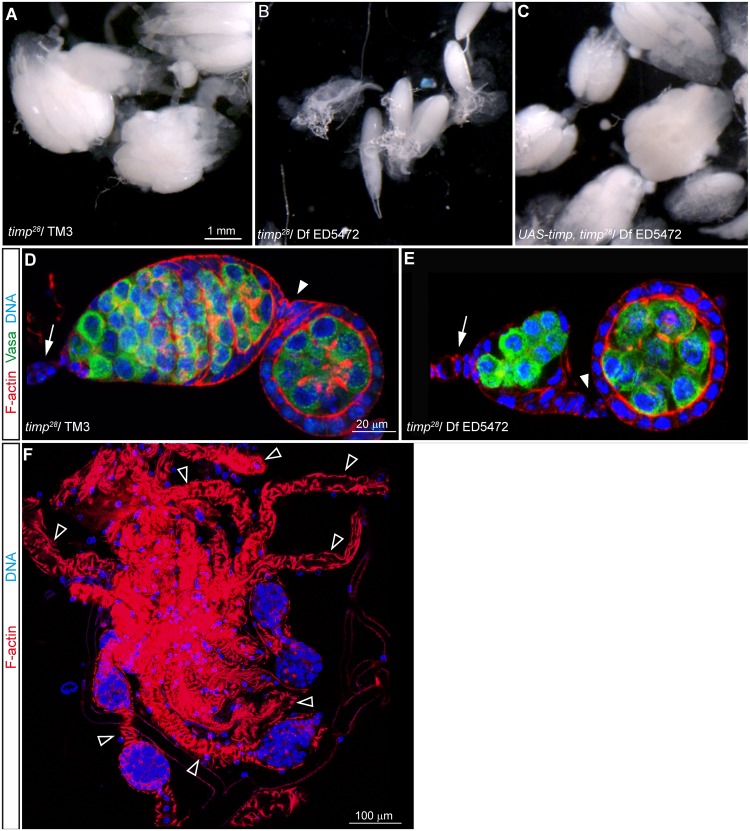
Fig 5. Ovaries from timp mutant females are smaller and lack integrity.
(A) Ovaries dissected from 2-week old control females. (B) Small disorganized ovaries dissected from a 2-week old timp null mutant. (C) Normal ovary morphology restored in ovaries dissected from 2-week old females lacking the endogenous timp gene but carrying a UASt-timp transgene. (D) Germarium from a 2-week old control ovary stained with Rhodamine-Phalloidin to visualize F-actin (red), Vasa to label de germline cells (green) and Hoescht to mark DNA (blue). (E) Germarium from a 2-week old mutant female stained as before. Note the marked decrease in the number of developing cysts within the germarium. (F) Ovary from a 4-week old mutant female stained to visualize the outline of the tissue and DNA. Empty arrowheads point to depleted ovarioles containing very few or no follicles. Arrows: terminal filament. Arrowheads: first interfollicular stalk. Images can be composites of several focal planes.

