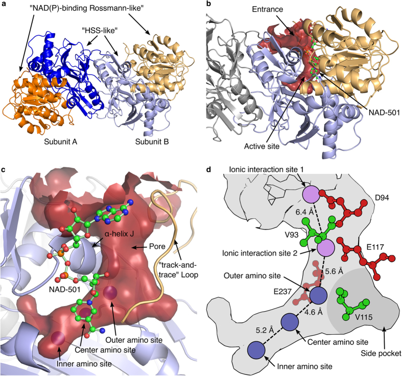
Figure 2. Overall structure of the homodimeric BvHSS.
(a) Cartoon representation of the BvHSS (PDB ID: 4PLP) dimer with domain 1 (“NAD(P)-binding Rossmann-like”) in orange (subunit A)/light orange (subunit B) and domain 2 (“homospermidine-synthase (HSS)-like”) in blue (subunit A)/light blue (subunit B). (b,c) Substrate binding pocket displayed as a surface-rendered cavity in red with adjacently bound NAD+ (in ball-and-stick representation). (c) Outer, center, and inner amino sites are indicated as blue spheres inside the red surface-rendered cavity, and the “track-and-trace” loop is given as a cartoon representation in light orange. (d) Representation of the substrate-binding pocket shown in (c). Amino sites are displayed as blue circles, and ionic interaction sites are displayed as violet circles. Amino acids providing the “ionic slide” (ball-and-stick representation) with acidic side chains are colored in red, and those with non-polar side chains are colored in green. The approximate position of the side pocket on the rear side of the binding pocket is indicated as a dark gray area.

