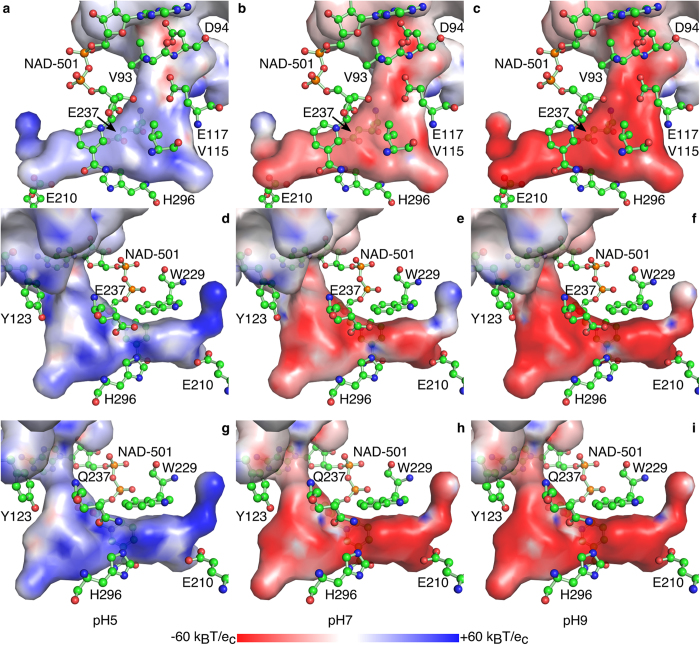
Figure 5. Representation of the electrostatic potential of the surface of the binding pocket at pH 5, 7, and 9.
The electrostatic potential at the surface of the substrate-binding pocket (compare with Fig. 2b,c) is represented as a color gradient from red (−60 kBT/ec) over white (0 kBT/ec) to blue (+60 kBT/ec) at pH 5 (a,d,g), pH 7 (b,e,h), and pH 9 (c,f,i) from two opposing orientations for BvHSS wild-type (a-f, PDB ID: 4PLP) and one orientation for BvHSS variant E237Q (g-i, PDB ID: 4XQG). The cofactor NAD+ and the important residues Val-93, Asp-94, Val-115, Glu-117 (“ionic slide”), Tyr-123, Glu-210, Trp-229, Glu-237, and His-296 are superimposed in ball-and-stick representation. Residues Glu-232, Glu-298, Tyr-323, and Tyr-325 are not shown for clarity, see Fig. 3 instead.

