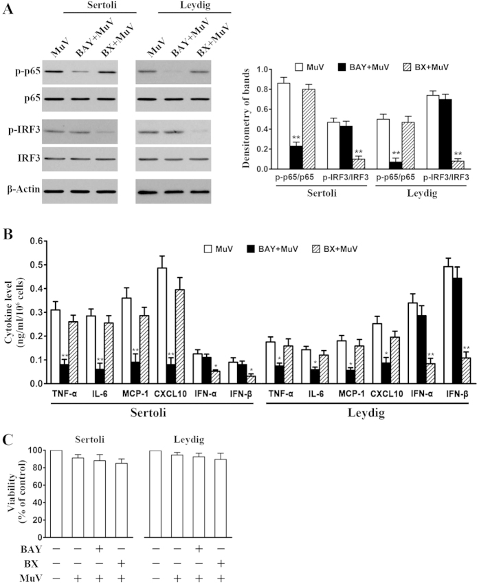
Figure 3. Involvement of NF-κB and IRF3 activation in cytokine expression.
(A) Inhibition of NF-κB and IRF3 activation. Sertoli and Leydig cells were infected with 5 MOI MuV alone, or with MuV after a 2-h pretreatment with 10 μM BAY11-7082 (BAY), or with MuV after a 2-h pretreatment with 1 μg/ml BX795 (BX). At 3 h post MuV infection, p-p65 and p-IRF3 levels were determined by Western blot (left panels). Densitometry of bands was analyzed with ImageJ software (right panel). (B) Effects of BAY11-7082 and BX795 on cytokine secretion. Sertoli and Leydig cells were treated as described in (A). The cytokine levels in the culture medium were measured with ELISA at 24 h post MuV infection. (C) Cell viability. Cells were treated as described in (A). Cell viability was assessed using an MTT assay at 24 h post MuV infection. Images represent at least three independent experiments. Data are presented as the means ± SEM of three experiments. *p < 0.05, **p < 0.01.

