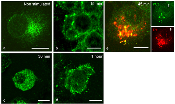
Figure 3. PC1/3 trafficking is affected by CpG-ODN internalization.
PC1/3 was labelled by indirect immunofluorescence using anti-PC1/3 (green, Alexa488) and imaged by confocal microscopy. (a) Non-stimulated NR8383 cells. (b–e) NR8383 cells incubated with 100 nM CpG-ODN 2006 for the indicated durations. (e) Co-localization of PC1/3 (green, Alexa488) and Texas Red labelled dextran identified PC1/3-positive vesicular structures as lysosomes (big) and endosomes (small). Bars = 10 μm.

