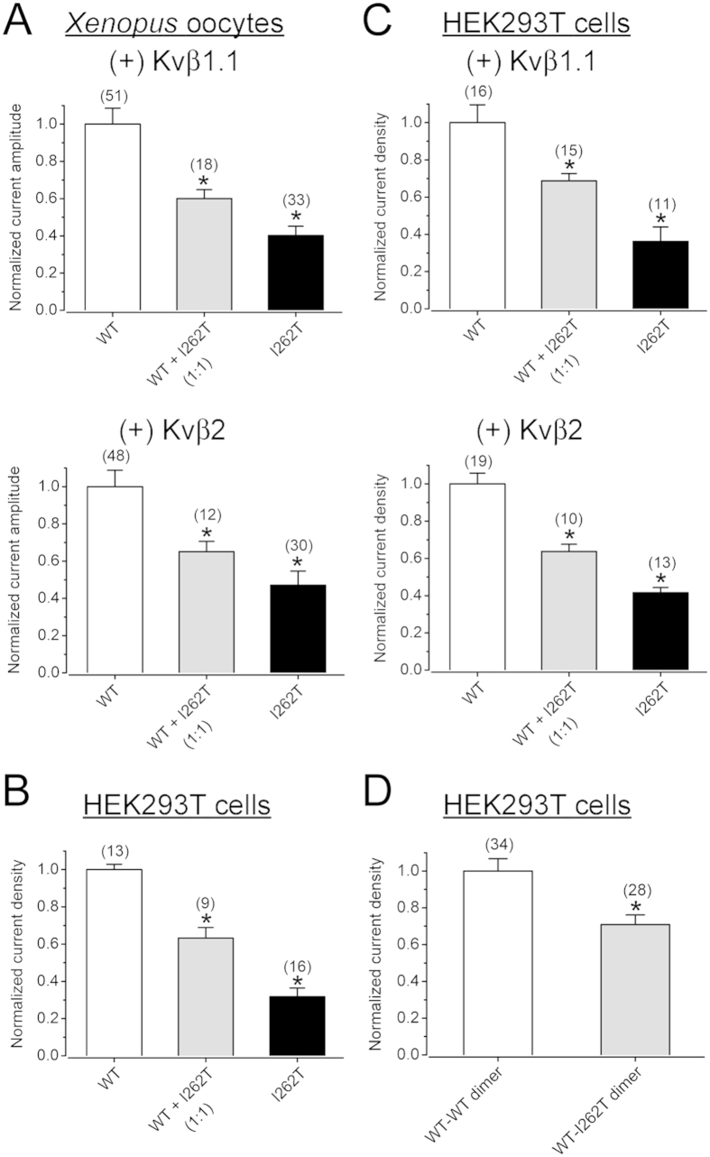
Figure 2. Co-expression with Kvβ subunits does not reverse the current suppression effect of I262T.
(A) Normalized peak current amplitudes (at + 60 mV) in Xenopus oocytes. Despite the presence of Kvβ1.1 or Kvβ2 subunits, 262T displays significant dominant-negative effects. (B,C) Normalized peak current densities (at + 60 mV) in HEK293T cells. I262T shows comparable current suppression effects in the absence or presence of Kvβ subunits. (D) Normalized peak current densities (at + 60 mV) of Kv1.1 WT-WT dimer and WT-I262T dimer in HEK293T cells. The voltage protocol is the same as that in Fig. 1A. Asterisks denote a significant difference from the corresponding WT control (*, t-test: p < 0.05). Kv1.1α and Kvβ subunits were co-expressed in the molar ratio 1:5. See Supplementary Figs S1 and S2 for representative current traces.

