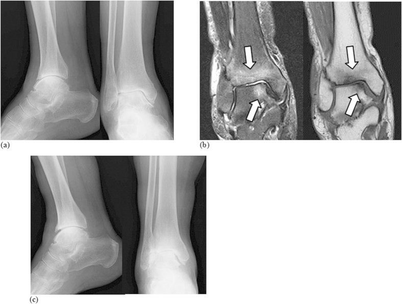
Figure 1.
(a) Frontal (right panel) and lateral (left panel) plain radiographs showed mild joint space narrowing and osteosclerotic changes (KL grade IV). (b) Frontal T1W (right panel) and STIR (left panel) MR images. Bone signal changes depicted as low intensity by T1W and high intensity by STIR images along the talocrural joint line were evident (arrows). (c) Frontal (right panel) and lateral (left panel) views of plain radiographs showed increased joint space narrowing and osteosclerotic changes (KL grade IV).

