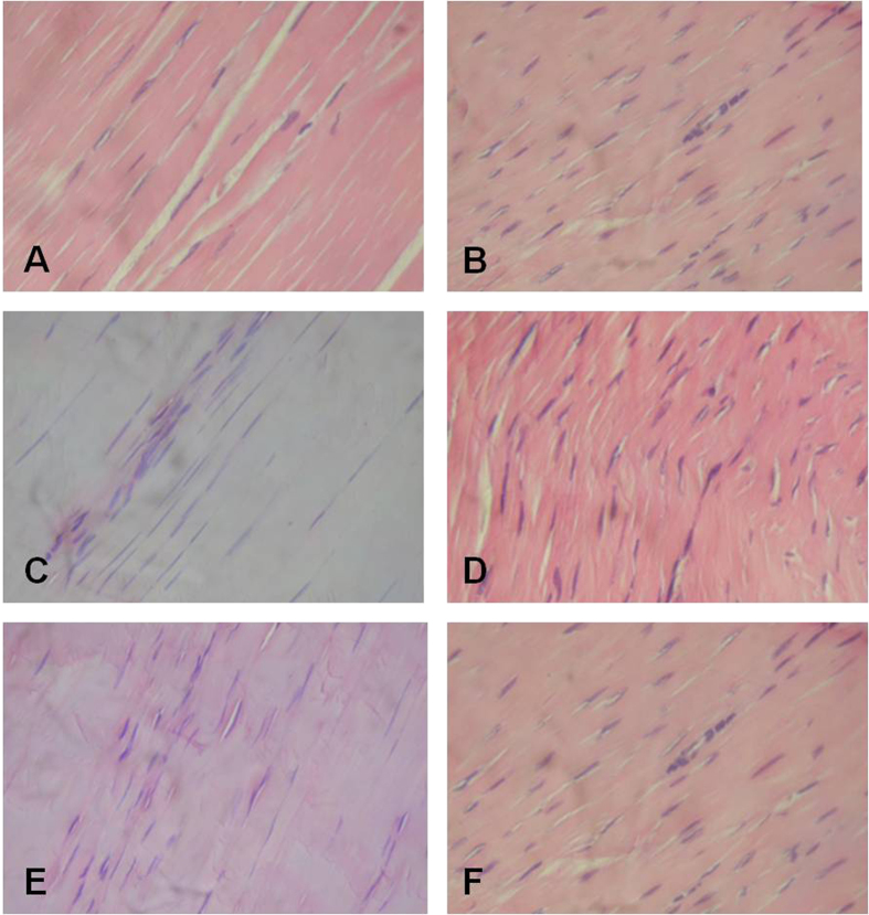
Figure 4. Tendon histology slides (H&E 40X).
(A) Longitudinal section of a healthy Achilles tendon showing normal parallel orientation of the collagen fibres and presence of tenocytes with characteristic elongated nuclei. (B) Longitudinal section Achilles tendon 10 days after collagenase treatment showing obvious changes in the orientation of the collagen fibres, increased tenocyte number with roundness of their nuclei. (C) Longitudinal section of the Achilles tendon treated with LR-PRP after 4 weeks, showing a slight increase in the tenocyte number. However, these cells predominately maintain the typical elongated structure of their nuclei. (D) Longitudinal section of the Achilles tendon treated with PBS after four weeks, showing a slight change in the orientation of the collagen fibers; the tenocytes are increased and some of them exhibit elongate or rounded nuclei. (E) Longitudinal section of the Achilles LR-PRP-treated after 12 weeks, the quantity of tenocytes is diminished and the shape of their nuclei is elongated. (F) Longitudinal section of the PBS-treated Achilles tendon after 12 weeks in a normal orientation of its collagen fibres and showing a decreased number of tenocytes that conserve the roundness of their nuclei.

