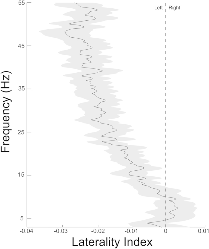
Figure 1. High-frequency resting oscillations are left-lateralized in children.

Linear regression of spectral power reveals cerebral asymmetry at high (20-50 Hz) but not low (3-7 Hz) frequencies. The extent of left asymmetry increases with increasing frequencies into the gamma range. The shaded region represents one standard error.
