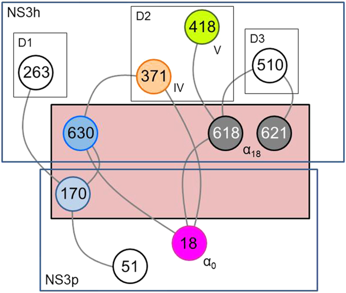
Figure 3. Schematic representation of NS3 coupled residue pairs.

Network of NS3 residues (nodes) with correlated mutations. The inter-domain interface is highlighted in salmon. Nodes are color coded to differentiate functional elements and highlight their role in NS3 protein structure and function33,34: magenta, light blue – peptide and MAVS cleavage; blue – natural protease substrate; grey, orange, green – molecular rearrangements and RNA replication. Annotation: D1 – helicase domain 1; D2 – helicase domain 2; D3 – helicase domain 3; IV – motif IV; V – motif V; α0 – helix α0; α18 – helix α18.
