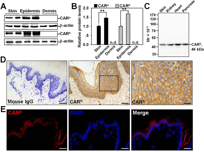
Figure 1. Expression and localization of CAR in normal human skin.
(A) Immunoblotting of CAR in lysates from normal human skin, epidermis, and dermis (~40 μg proteins/lane) with β-actin serving as a loading control. Two antibodies were utilized, anti-CARa is a rabbit origin antibody, while anti-CARb is a mouse origin antibody that was used in all following experiments. (B) The CAR level in skin after normalization against its corresponding β-actin was arbitrarily set at 1. Error bars represent means ± SD from four individuals (n = 4); **p < 0.01; n.d., not detectable. (C) Specificity of the mouse anti-CARb antibody was demonstrated by an immunoblot using normal human skin, kidney, heart, and pancreas lysates. (D) Immunohistochemistry staining of CAR on normal human skin paraffin section, in which the positive CAR staining appeared as brownish precipitates (middle and right images). Image on the right was the enlargement of the boxed area in the middle image. Sections incubated with normal mouse IgG instead of the mouse anti-CAR served as negative control (left). Scale bars: left, 50 μm; middle, 30 μm; right, 10 μm. (E) Immunohistofluorescent staining of CAR (red) on normal human skin paraffin section. DAPI was used for nuclear staining. Scale bar: 50 μm.

