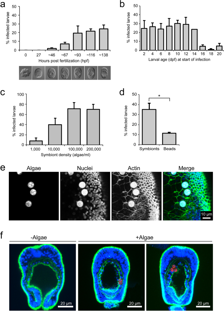
Figure 3. Symbiosis establishment during larval development.
(a) Quantification of symbiont uptake efficiencies in Aiptasia embryos and early planula larvae: early (<32-cell-stage) embryos were exposed to a constant environmental supply of Symbiodinium strain SSB0121 (10,000 algae/ml) and subsets were sampled at the times indicated (hpf) to assess infection efficiency. Representative DIC images are shown below each timepoint. Error bars are SEM, n = 3 replicate experiments. (b) Quantification of symbiont uptake efficiencies for larvae between 2 and 20 dpf: larvae at the ages indicated (dpf) were incubated with Symbiodinium strain SSB01 (10,000 algae/ml) for four days, after which infection efficiency was assessed. Error bars are SEM, n = 3 replicate experiments. (c) Quantification of symbiont uptake efficiencies after four days exposure for larvae 6–7 dpf at increasing algal concentrations. Error bars are SEM, n = 3 replicate experiments. (d) Quantification of comparison of uptake efficiency between SSB01 algae and inert fluorescent beads for larvae 4 dpf after four days exposure. Error bars are SEM, ***p < 0.001 as determined by Student’s t-test for unpaired data, n = 3 replicate experiments. (e) Representative fluorescence microscopy images of phagocytosed symbionts in endodermal cells of larvae 8 dpf. Hoechst-stained nuclei are shown in blue, phalloidin-stained F-actin to mark cell outlines in green, and endogenous autofluorescence of algal chlorophyll in red. Note that algae exhibit strong autofluorescence in all channels. (f) Representative confocal microscopy images of larvae 10 dpf with or without symbionts. Fluorescence channels are as in (e).

