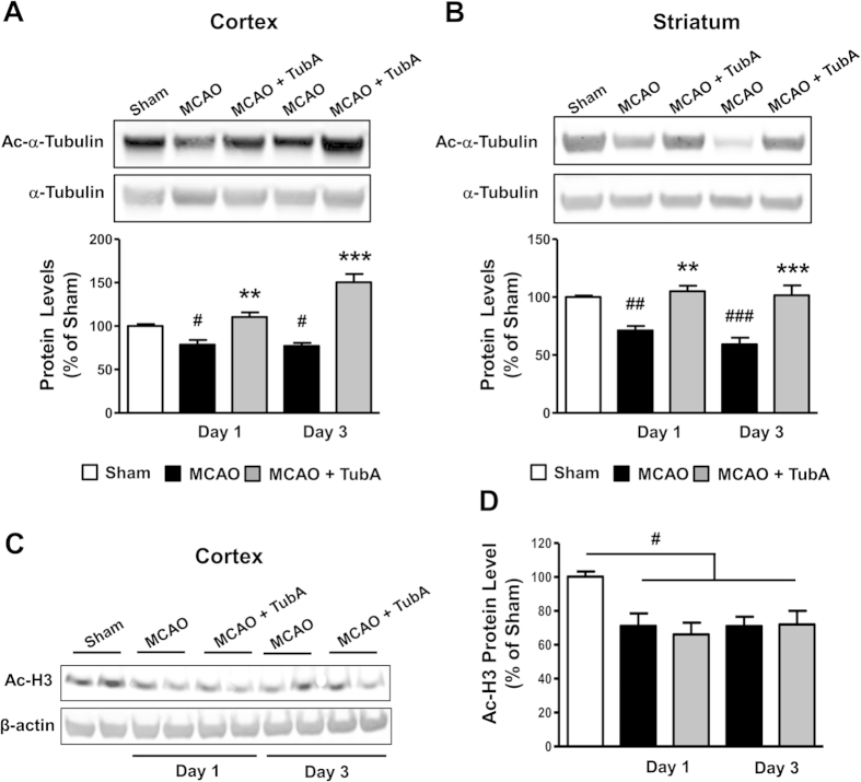
Figure 4. TubA (25 mg/kg) restored α-tubulin acetylation levels in the ischemic cortex and striatum on Days 1 and 3 after ischemia.
Middle cerebral artery occlusion (MCAO) reduced the levels of acetylated α-tubulin (Ac-α-Tubulin) in the ischemic cortex (A) and striatum (B) on Days 1 and 3 after ischemia. TubA significantly increased α-tubulin acetylation levels. (C) TubA had no effect on restoring acetylated histone H3 (Ac-H3) protein levels in the cortex on Days 1 and 3 after ischemia. Quantified data is shown in (D). #P < 0.05, ##P < 0.01, ###P < 0.001 compared with sham control; **P < 0.01, ***P < 0.001 compared with MCAO group; n = 6 per group (for α-tubulin acetylation); n = 4 per group (for histone H3 acetylation).

