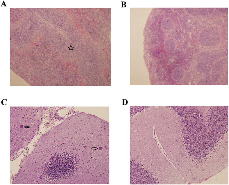
Figure 1. Histopathological lesions in mouse tissues infected with Toxoplasma gondii Pru strain (10 cysts per mouse by oral route, H&E stain).
(A) A section of spleen from infected mice at 14 DPI. Pentagram indicates the largely scattered white pulp; (B) A section of spleen from normal mice at 14 DPI showing no histological abnormalities; (C) Brain histology of infected mice at 21 DPI. Arrow indicates the tissue cyst; (D) Brain histology of normal mice at 21 DPI without any pathological changes. Magnifications: 40X (A,B); 100X (C,D).

