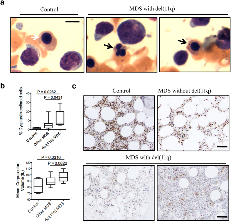
Figure 7. MDS patients with H2AX deficiency exhibit increased dyserythropoiesis and poor prognosis.
(a) Wright-Giemsa staining of representative bone marrow smears from patients. White arrow indicates a normal orthochromatic erythroblast, and black arrows indicate a dysplastic orthochromatic erythroblast (middle) and an erythroblast with a Howell-Jolly body (right). Scale bar: 10 μm. (b) Quantification of dysplastic erythroblasts. Control = bone marrow smears from lymphoma-stage patients, n = 5; Other MDS = bone marrow smears from MDS patients without del(11q), n = 17; del(11q) MDS = bone marrow smears from MDS patients with del(11q), n = 7. (c) Immunohistochemical staining for H2AX in bone marrow core biopsies from patients. Scale bar: 100 μm. Control: representative of three lymphoma-stage patients; MDS without del(11q): representative of eight patients; MDS with del(11q): representative of all seven patients. Immunohistochemical staining was performed for all patients, and staining representative of patients with similar results is presented.

