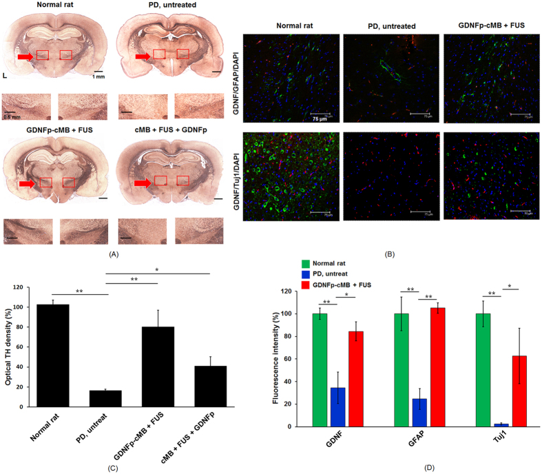
Figure 5. Immunohistochemical fluorescence staining.
(A) Representative photomicrographs of SN sections immunostained for TH from normal rat, PD rat, GDNFp-cMB+FUS group, and cMB+FUS+GDNFp group. (B) IHC distribution of GDNF, GFAP, and Tuj1 expression in the sonication region after gene transduction. Transfected GDNF/GFAP/DAPI (top) and GDNF/Tuj1/DAPI (bottom) for the normal rat, PD rat, and PD GDNFp-cMB+FUS group, respectively. GDNF: red; Tuj1: green; GFAP: green; DAPI: blue. (C) The optical density of TH fibers in the lesioned hemisphere compared to the non-lesioned hemisphere. (D) The fluorescence intensity of GDNF, GFAP and Tuj1 in the lesioned site compared to the normal rat. Single asterisk, p < 0.05; double asterisk, p < 0.01. Data were analyzes by one-way ANOVA (post hoc test: Dunnett; degrees of freedom: 8, 6; F value: 46.9, 39.2) and presented as mean ± SEM (n = 6 per group). All of the data were compared with PD untreated rats.

