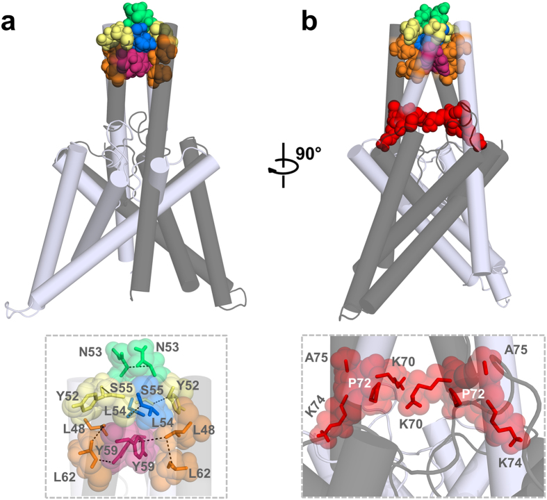
Figure 6. Hydrophobic interactions stabilize the tip of the TASK-1 cap structure.
TASK-1 ‘hits’ identified in the alanine scan are highlighted in the TASK-1 homology model based on the domain-swapped TREK-2, following a 100 ns MD simulation. The two different subunits are shown in gray and light gray. (a) Residues stabilizing the tip of the cap structure are shown in space fill mode and a zoom-in is provided at the bottom. (b) 90° rotation of the model. Mutations resulting in a loss of ‘conductivity’ are illustrated in red space fill and a zoom-in showing these residues located in the EIP is provided at the bottom.

