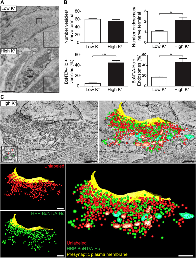
Figure 1. BoNT/A-Hc is internalized in a subpopulation of synaptic vesicles in presynaptic hippocampal nerve terminals.
Hippocampal neurons were incubated with HRP-BoNT/A-Hc (10 μg/ml) for 5 min in low K+ or high K+ buffer, fixed and processed for electron microscopy. (A) Representative images of HRP-BoNT/A-Hc endocytosis into control or high K+-treated presynaptic regions (50 nm sections). Scale 500 nm. (B) The total number of vesicles (40–80 nm diameter) and endosomes (>80 nm) in synaptic regions were quantified, together with the percentage of compartments containing HRP-BoNT/A-Hc. Mean ± sem of 4–6 independent neuron preparations (>40 synapses analyzed/preparation). **p < 0.01, ***p < 0.001. (C) An optical slice and three-dimensional reconstruction of a representative presynaptic region that has endocytosed HRP-BoNT/A-Hc (green asterisks in enlargement). HRP-BoNT/A-Hc-containing organelles are distributed randomly in relation to non-labeled compartments and the presynaptic plasma membrane. Scale 200 nm.

