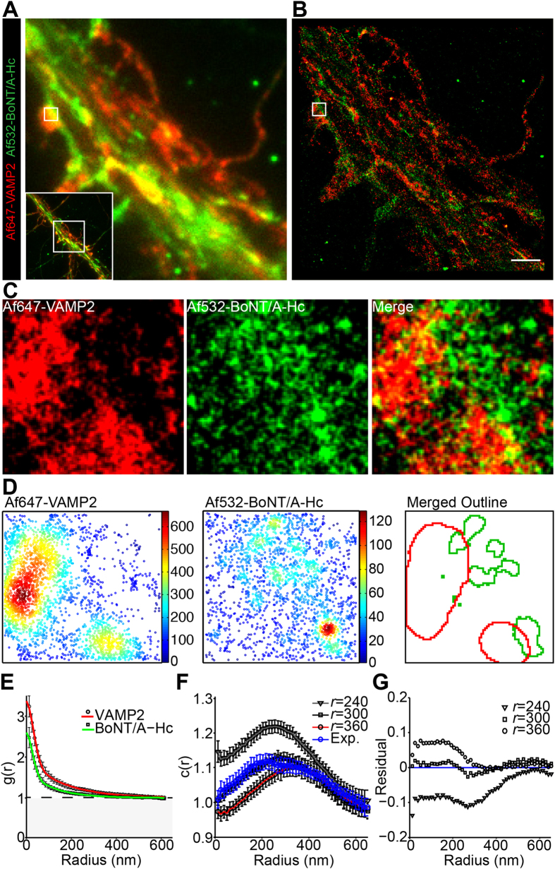
Figure 2. Segregation of Af532-BoNT/A-Hc clusters from VAMP2 in hippocampal nerve terminals.
Hippocampal neurons were incubated with Af532-BoNT/A-Hc (150 nM) for 5 min in high K+ buffer, fixed and immunolabeled using a VAMP2 antibody, prior to GSDIM imaging and processing. A representative low-resolution image (A) and corresponding reconstructed super-resolution localization image (B) are shown. Scale 2 μm. (C) Enlargement of the synaptic region indicated in (B). (D) Cluster maps generated from Ripley’s K-function and outlines of interpolated maps are shown for the enlargement. (E) Mean (±sem) auto-correlation functions of each channel fitted using equation (4) (21 regions of interest (ROIs) from 2 independent experiments). (F) Cross-correlation function of Af647-VAMP2 and Af532-BoNT/A-Hc at VAMP2-enriched synaptic regions (Exp.) (21 regions of interest from 2 independent experiments). Cross-correlation functions were also used to analyze simulated data of clusters that were separated by varying radii (indicated by r). (G) Residuals were calculated by subtracting the cross-correlation values of simulated data from the experimental data.

