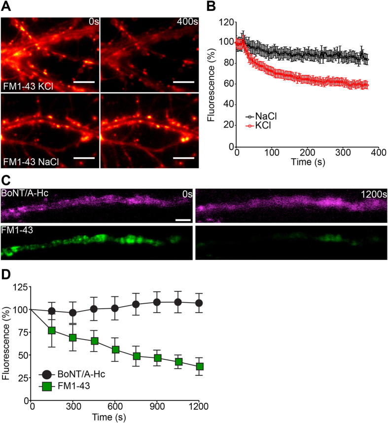
Figure 6. BoNT/A-Hc is retained in non-releasable vesicles upon a secondary stimulus.
(A,B) Cultured hippocampal neurons were incubated with FM1-43 (4 μM) for 2 min in the presence of high K+ and following a 12–15 min recovery were subjected to a secondary stimulus of KCl (30 mM) or NaCl (30 mM). Representative before and after images are shown (A). (B) Quantification of a representative experiment showing the change in fluorescence during stimulation. Data are plotted as mean ± sem (n = 32–35 nerve terminals from a representative experiment). Amphibian neuromuscular junction preparations were incubated with Atto647N-BoNT/A-Hc (800 nM) for 20 min in the presence of high K+ Ringer’s solution. FM1-43 (5 μM) was applied in the last 3 min. Preparations were washed with low K+ Ringer’s solution and left to recover for 20 min. Destaining was induced by replacing the solution with high K+ Ringer’s solution and confocal time-lapse imaging of nerve terminals was carried out. (C) Representative time-lapse images of a nerve terminal showing destaining of FM1-43 but not Atto647N-BoNT-A-Hc in response to the secondary high K+ stimulus. Scale 10 μm. (D) Rate of destaining of Atto647N-BoNT-A-Hc (magenta) and FM1-43 (green) in response to the secondary high K+ stimulus (n = 3 independent preparations).

