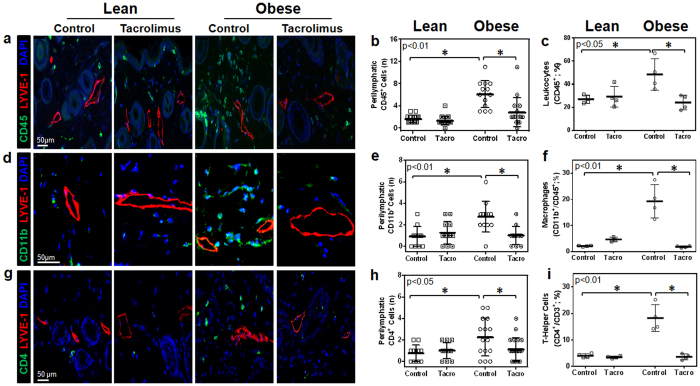
Figure 2. Topical tacrolimus decreases perilymphatic inflammation in obese mice.
(a) Representative high power photomicrographs (40x) and quantification of (b) perilymphatic inflammation in hindlimb sections stained for CD45 (green) and LYVE-1 (red; n = 5 animals with 4–5 HPF/animal, p < 0.01). (c) Hindlimb tissues assessed using flow cytometry to identify CD45+ cells as a percentage of live cells (p < 0.05). (d) Representative high power photomicrographs (80x) and quantification of (e) perilymphatic inflammation in hindlimb sections stained for CD11b (green) and LYVE-1 (red; n = 5 animals with 4–5 HPF/animal, p < 0.01). (f) Hindlimb tissues assessed using flow cytometry to identify macrophages (CD11b+/CD45+) as a percentage of total live cells (n = 4 animals/group, p < 0.01). (g) Representative high power photomicrographs (40x) and quantification of (h) perilymphatic inflammation in dermal hindlimb sections stained for CD4 (green) and LYVE-1 (red; n = 5 animals with 4–5 HPF/animal, p < 0.05). (i) Hindlimb tissues assessed using flow cytometry to identify T helper cells (CD4+/CD3+/CD45+) as a percentage of total live cells (p < 0.01).

