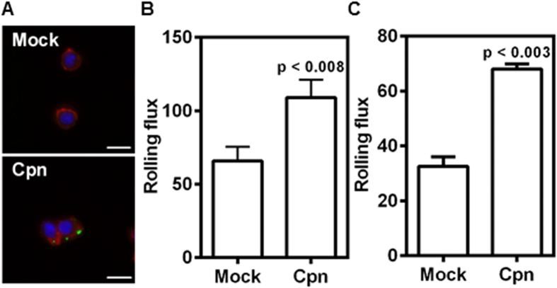
Figure 1. C. pneumoniae infection increases monocyte recruitment to E-selectin and endothelium under flow.
Monocytes were infected with mock PBS or Chlamydial EB (MOI 1) for 8 h. (A) The cells were stained for Chlamydia pneumoniae (green), actin (red), and nucleus (blue). Representative image of Mock and Cpn infected cells are shown (Scale bar, 10 μm). (B,C) In another experiment the cells were re-suspended at a concentration of 0.5 million/ml in media and perfused at 1 dyn/cm2 over micro-channels coated with E-selectin (B); and over confluent aortic endothelium activated for 4 h with 20 ng/ml of TNFα (C). The number of cells rolling on the surface was obtained from video microscopy (20 fps) for 5 minutes. The results are mean ± SEM of one representative experiment performed in triplicate, and the experiments were repeated five times. The statistical significance in the parameters between the groups was shown as p value from t test.

