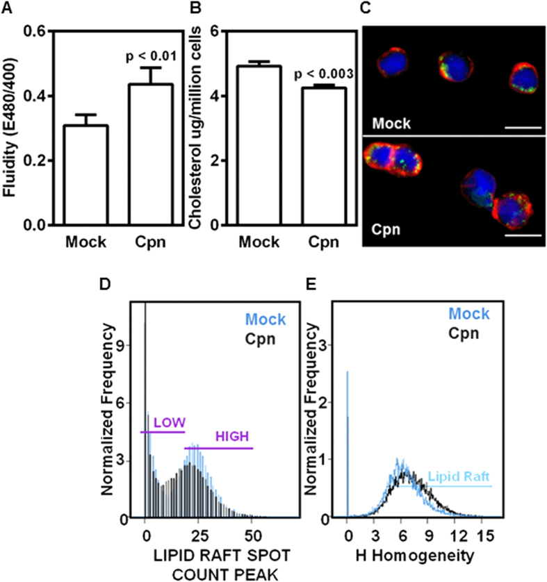
Figure 3. C. pneumoniae infection increases membrane fluidity and uniformity of lipid raft distribution.
Uninfected (mock) or infected (Cpn) monocytes were analyzed for fluidity of membrane using a fluorescence plate reader and the results were plotted as ratio of emissions at 480 and 400 nm (A); analyzed for total cholesterol using fluorescence plate reader (B); stained for membrane lipid rafts (green), actin (red), and nucleus (blue), and visualized by confocal microscopy (Scale bar, 10 μm) (C). The results are mean ± SEM of five different experiments performed in triplicate. The statistical significance in the parameters between the groups was shown as p value from t test. In another experiment monocytes were stained for lipid rafts and analyzed for lipid raft distribution by spot count analysis (D); and homogeneity (E) by imaging flow cytometry. The results are from one representative experiment performed in triplicate, and all the experiments were repeated five times.

