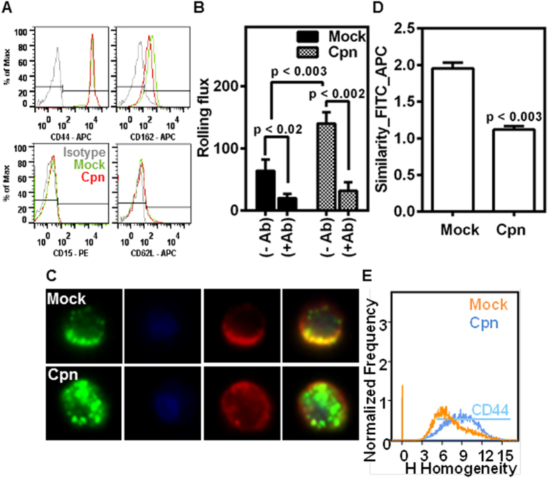
Figure 4. CD44 mediates rolling and is more uniformly distributed in infected cells.
Uninfected (mock) or infected (Cpn) monocytes were stained with CD44, CD15, CD162, or CD62L antibodies, and analyzed by flow cytometry (A). In another experiment, the cells (mock or Cpn) after 8 h of infection were blocked with 0 or 1 μg of CD44 antibody per million cells for 30 minutes, washed and re-suspended at a concentration of 0.5 million/ml in media and perfused on activated endothelium at 1 dyn/cm2, and the rolling interactions were quantified by video microscopy (B). (C–E) Cells after 8 h of infection were stained for CD44 (red), lipid raft (green) and nucleus (blue) and visualized by imaging flow cytometry, with representative images (C); co-localization of CD44 and lipid rafts (D); and homogeneity in the distribution of CD44 (E) are shown. The results are mean ± SEM (B,D) of one representative experiment performed in triplicate, and all the experiments were repeated three times. The statistical significance in the parameters between the groups was shown as p value from ANOVA (B) or t test (D).

