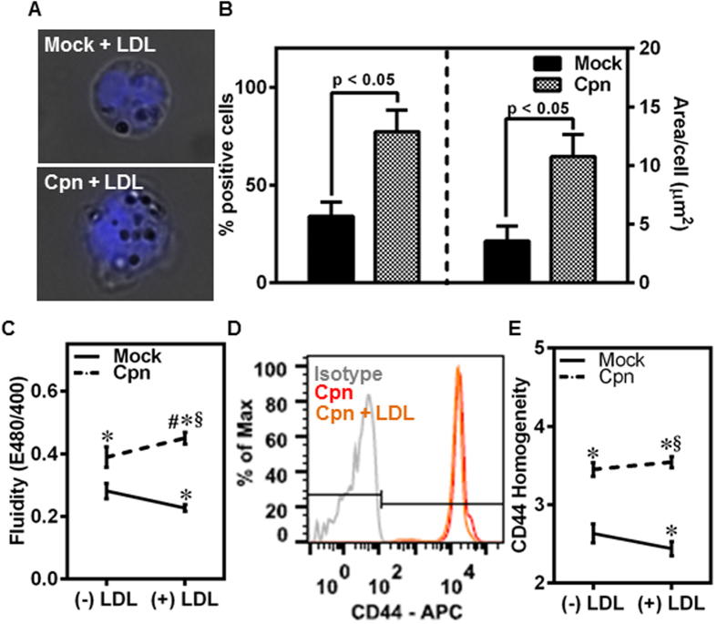
Figure 5. LDL increases CD44 homogeneity in infected, but not uninfected, monocytes.
(A–E) Monocytes were infected with mock PBS or Chlamydial EB (MOI 1) for 4 h and further incubated for 4 h with 0 or 100 μg/ml of LDL. The cells were stained for: LDL with Oil red O (droplets) and nucleus (blue) (A); quantitative uptake of LDL using ImagePro software (B); membrane fluidity estimation using fluorescent plate reader (C); CD44 expression assayed by flow cytometry (D); and CD44 distribution on the membrane by imaging flow cytometry (E). The results are mean ± SEM (B,C,E) of three different experiments performed in triplicate. The statistical significance in the parameters between the groups was shown as p value from t test (B). The *, # and § denote statistically significant change in the parameters of the groups in comparison to mock, Cpn and mock + LDL respectively (p < 0.05, ANOVA) (C,E).

