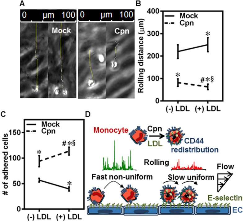
Figure 7. LDL increases monocyte rolling and adhesion to endothelium.
(A–C) Monocytes were infected with mock PBS or Chlamydial EB (MOI 1) for 4 h and further incubated for 4 h with 0 or 100 μg/ml of LDL. The cells were then re-suspended at a concentration of 0.5 million/ml in media and perfused on activated aortic endothelium at 1 dyn/cm2, and the interactions were visualized by video microscopy. Micrograph of tracks for uninfected (Mock) and infected (Cpn) conditions (A); distance rolled by the cells before adhering firmly (B); and number of cells firmly adherent on endothelium under various conditions (C). The results are mean ± SEM of three different experiments performed in triplicate. The *, # and § denote statistically significant change in the parameters of the groups in comparison to mock, Cpn and mock + LDL respectively (p < 0.05, ANOVA). (D) Proposed biophysical mechanism for the increased adhesion of infected monocytes to the endothelium in the presence of LDL.

