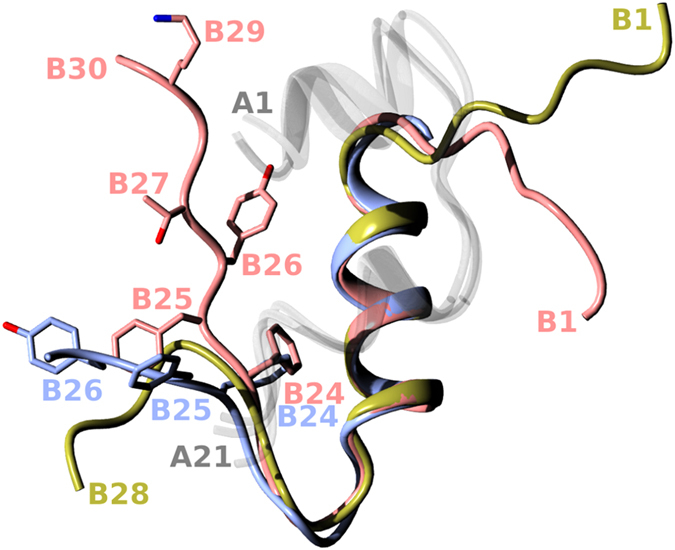
Figure 1. An overlay of human insulin structures that were key templates in this work.

All A chains (not relevant here) are in a translucent white; the wild-type B-chain (pdb 1mso) is in coral; the B-chain insulin structure attained in the complex with IR (pdb 4oga) is in blue; and the B-chain of one of the representative B26-turn-containing analogues (AsnB26-insulin, pdb 4 ung) is in gold.
