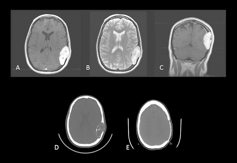
Fig. 2.

Case 2. Preoperative axial postcontrast T1-weighted MRI (A), axial T2-weighted MRI (B), and coronal postcontrast T1-weighted MRI reveal a large tumor of the parietal bone with its epicenter in the diploe of the skull. The typical pattern of inhomogeneous enhancement on T1-weighted imaging and hyperintensity on T2-weighted imaging is again seen. Preoperative (D) and postoperative (E) axial CT scans reveal complete tumor removal with custom cranial implant replacing the removed bone.
