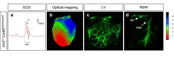
Figure 2. Loss of Cx40 results in abnormal electrical activation of the ventricles without morphological defects of the VCS.
(a) Representative surface ECG trace shows QRS prolongation and notched R’ wave in Irx3+/+;Cx40EGFP/EGFP mice. (b) Optical mapping in Irx3+/+;Cx40EGFP/EGFP heart exhibited RBB block, similar to Irx3−/− heart. (c,d) VCS structures visualized by Cx40-EGFP in both LV (c) and RSW (d) are normal in Irx3+/+;Cx40EGFP/EGFP mice. LV, left ventricle; RSW, right ventricular septal wall; HB, his-bundle; RBB, right bundle branch.

