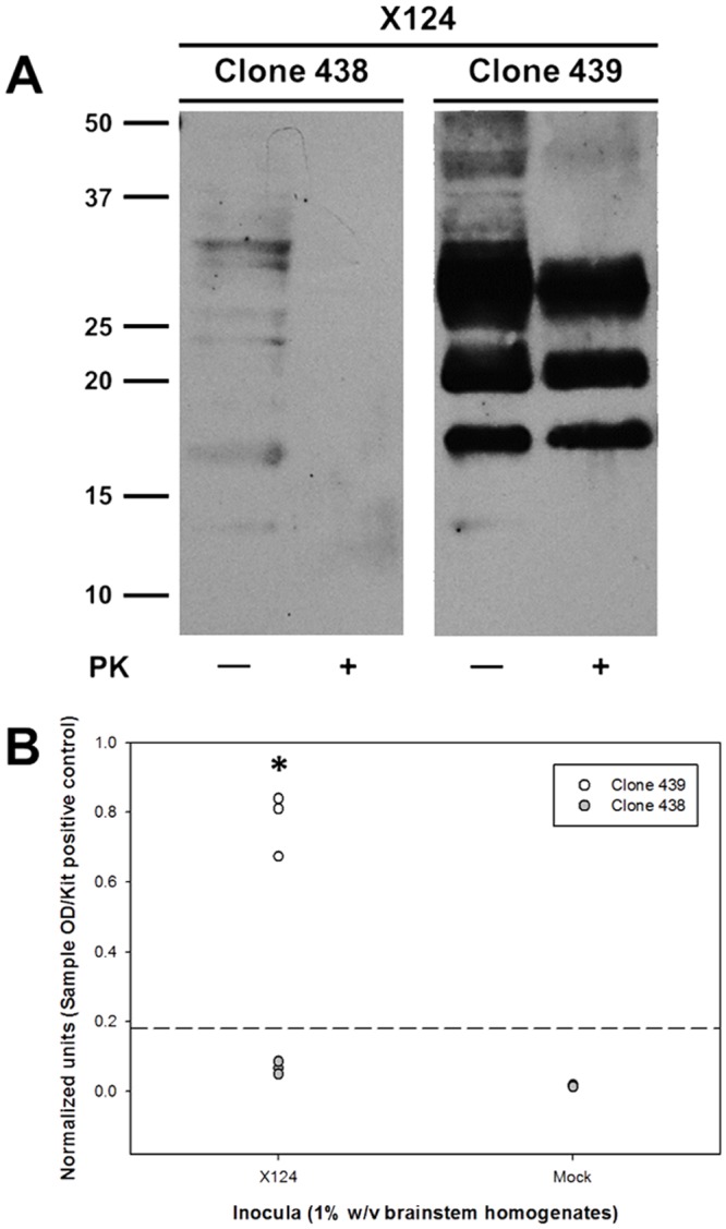
Fig 1. Characterization of differential prion susceptibility in ovine microglia clones.

Microglia clones were inoculated with 1% (w/v) brainstem homogenates from either scrapie-positive (“X124”) or scrapie-naïve (“Mock”) sheep. Inoculated cells were passaged on a weekly basis and then tested for the accumulation of nascent PrPSc at passage three by immunoblotting (A) and at passage four by ELISA (B). Immunoblot picture (A) depicts results from one culture replicate inoculated with scrapie-positive brain homogenates and is representative of three independent experiments. In graph (B), each circle represents the mean of three culture replicates from each of three independent experiments (i.e., total of six circles per treatment, three for 439 and three for 438), and the dashed line indicates the assay cut-off threshold for detection of PrPSc. Values of normalized units in the X124 group for clone 439 are significantly higher than those of in clone 438 (*: P = 0.0048, unpaired t-test). Values of normalized units between X124 and mock groups are statistically significantly different for the clone 439 (P = 0.0044, paired t-test) and clone 438 (P = 0.0065, paired t-test); however, the values for clone 438 fail to rise above the cut-off threshold and are negative by immunoblotting ([A] and S1 Fig).
