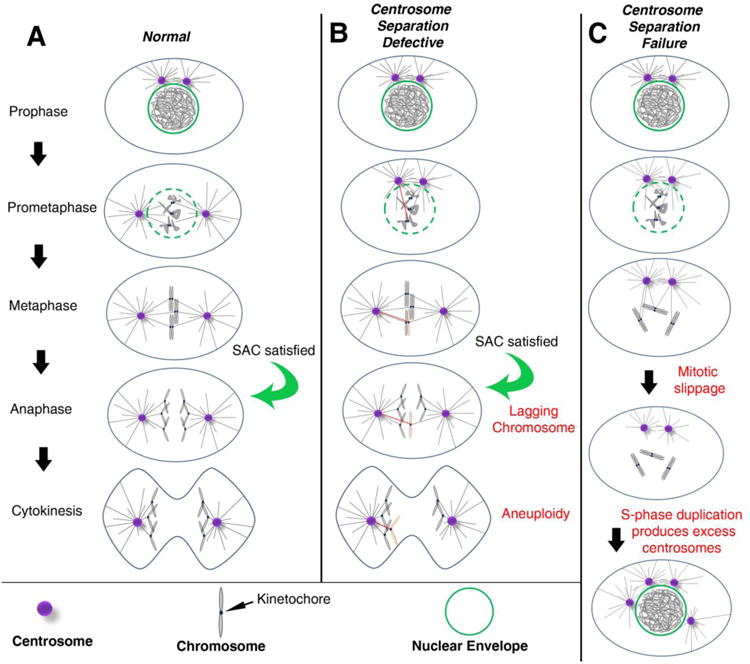
Figure 1. Centrosomes promote efficient bipolar spindle assembly.

(A) In most cell types, the pair of centrosomes (purple) is closely associated at the entry into mitosis (Prophase). During prophase and prometaphase, centrosomes separate from one another and take up residence on opposite sides of the nucleus. In prometaphase, the nuclear envelope (green) breaks down, allowing MTs (black lines) emanating from the centrosomes to enter the nuclear space, find the kinetochores, and ultimately establish amphitelic attachments (i.e. the kinetochores of sister chromatids are individually attached to MTs from opposing spindle poles). The chromosomes will then congress to the metaphase plate. Once all kinetochores are attached to MTs, the SAC is satisfied, providing the “go anaphase” signal. During anaphase, sister chromatids are segregated towards opposite poles of the spindle. Finally, during telophase, the cytokinetic furrow ingresses, dividing the cell into two daughter cells with identical chromosome complements (abscission and daughter cell formation not depicted). (B) When there are moderate early defects in centrosome separation, the centrosomes may remain closely apposed as the cell enters prometaphase. This is believed to favor incorrect attachments of MTs from both poles to the kinetochore of the same chromatid. The red MT indicates the inappropriate attachment to the affected chromosome (pink). Even in the presence of these merotelic attachments, the SAC can be satisfied, allowing anaphase onset. The opposing forces of MTs from opposite poles on the same kinetochore can result in a lagging chromosome, which, in some cases, will mis-segregate into the wrong daughter cell, resulting in aneuploidy. (C) When centrosomes fail to separate, MTs may not attach to some kinetochores, leaving the SAC unsatisfied. Eventually, mitotic slippage may allow for mitotic exit into G1 without chromosome segregation or cytokinesis, resulting in a tetraploid cell with both centrosomes. In the subsequent S-phase, both centrosomes duplicate, resulting in extra centrosomes.
