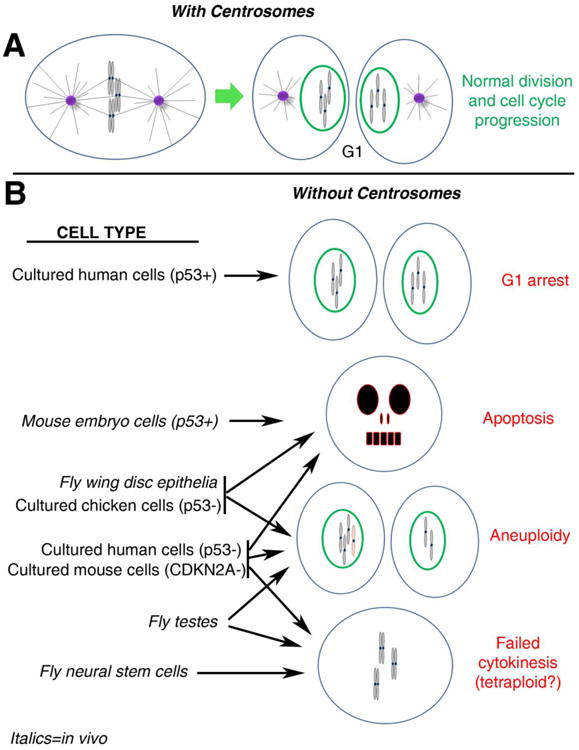
Figure 2. Differential effects of centrosome loss among model systems.

(A) In normal cells with centrosomes, a metaphase cell progresses through cytokinesis (not shown) to produce 2 daughter cells in G1, each with a normal chromosome complement and a single centrosome. To help visualize the consequences of chromosome segregation errors (e.g., aneuploidy), G1 chromosomes are depicted as condensed in this figure, though they would actually decondense upon mitotic exit. (B) The outcomes of mitosis in different cell types depleted of centrosomes are shown. p53 status is indicated for some relevant cell types (if not indicated, p53 is normal). Italics denote cells from in vivo studies. See text for more thorough descriptions of these studies. Not represented is the potential connection between mitotic error and accumulation of DNA damage; as observed in fly wing disc epithelia and cultured chicken cells. Note that lack of a connection to a specific outcome for a particular cell type does not necessarily indicate the outcome does not occur for that cell type. Rather it may simply have not been examined or reported in the relevant studies.
