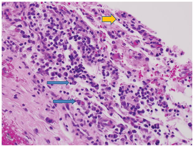
Figure 2.
High-power photomicrograph (hematoxylin-eosin, magnification ×125) demonstrating a membrane adjacent to a femoral component of a cobalt-chromium alloy in a patient with painful, persistent synovitis after total knee arthroplasty. Chronic inflammation is present, and the predominant cells are lymphocytes (yellow arrow) and plasma cells (blue arrows). No multinuclear giant cells and no polyethylene fragments are evident. The synovial biopsies showed the same pattern. (Courtesy of Maureen Bauer, MD, Durham, NC.)

