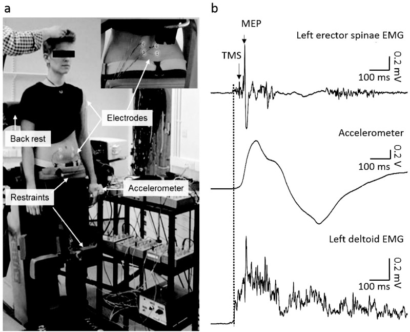
Fig 1. Experimental setup.
(a) Participants stood upright on a restraining device with pelvis and knees securely fixed to minimise movement of pelvis and lower limbs. Electrodes were attached over erector spinae at the 4th lumbar vertebral level (panel in top right), rectus abdominis and left deltoid. An accelerometer was positioned on the dorsum of the hand contralateral to the stimulation. (b) Representative data from a single subject showing left erector spinae EMG, left deltoid EMG and accelerometer data during dynamic shoulder flexion task. TMS was delivered 25 ms after the onset of deltoid EMG (dotted vertical line).

